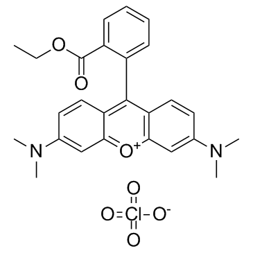
TMRE (Tetramethylrhodamine ethyl ester perchlorate)
A fluorogenic dye
此产品仅用于科学研究,我们不为任何个人用途提供产品和服务
-
包装价格促销价数量
-
5mg¥637.00510.00- +
-
10mg¥975.00780.00- +
-
25mg¥1937.001550.00- +
- 货号: ajce44348
- CAS: 115532-52-0
- 别名: 四甲基罗丹明乙酯; Tetramethylrhodamine ethyl ester perchlorate
- 分子式: C26H27ClN2O7
- 分子量: 514.95
- 纯度: >98%
- 溶解度: DMSO : ≥ 150 mg/mL (291.29 mM)
- 储存: Store at -20°C
- 库存: 现货
Background
Tetramethylrhodamine ethyl ester (TMRE) (perchlorate) is a non-cytotoxic cell-permeant fluorogenic dye most commonly used to assess mitochondrial function using live cell fluorescence microscopy and flow cytometry.1,2 It displays excitation/emission spectra of 550/575 nm, respectively. Due to the polarization of the mitochondrial membrane, TMRE is taken up into healthy mitochondria. However, when the membrane is depolarized, as in apoptosis, it is not taken up or is released from the mitochondria. Thus, the strength of the fluorescence signal in mitochondria is used to assess cell viability.
1.Farkas, D.L., Wei, M.-d., Febbroriello, P., et al.Simultaneous imaging of cell and mitochondrial membrane potentialsBiophys J.56(6)1053-1069(1989) 2.Sunaga, D., Tanno, M., Kuno, A., et al.Accelerated recovery of mitochondrial membrane potential by GSK-3β inactivation affords cardiomyocytes protection from oxidant-induced necrosisPLoS One9(11)e112529(2014)
Protocol
|
Cell experiment: |
The entire experiment should be performed at room temperature because temperature will directly impact mitochondrial transmembrane potential and TMRE staining. Cells should never be placed, centrifuged, incubated, or washed at 4°C or have ice-cold buffers or media added. Treat the cells with a cytotoxic stimulus. Harvest cells and resuspend at 5×10⁵ cells/mL in culture medium containing 150 nM TMRE. Incubate for 5 min at room temperature in the dark. Add Carbonyl cyanide 4-(trifluoromethoxy)phenylhydrazone (FCCP) (5 μM final concentration) to an aliquot of untreated cells and incubate for 5 min at room temperature in the dark. Turn on the appropriate laser on the flow cytometer. Set up a histogram plot to detect TMRE using log scale[1]. |
|
参考文献: [1]. Crowley LC, et al. Measuring Mitochondrial Transmembrane Potential by TMRE Staining. Cold Spring Harb Protoc. 2016 Dec 1;2016(12):pdb.prot087361. |
|
-

LX1606 Hippurate (Telotristat etiprate)
¥580.00 ¥725.00


没有评价数据